- J-STAGE home
- /
- Circulation Journal
- /
- Volume 84 (2020) Issue 11
- /
- Article overview
-
Chisato Izumi
Department of Cardiovascular Medicine, National Cerebral and Cardiovascular Center [Japan]
-
Kiyoyuki Eishi
Division of Cardiovascular Surgery, Nagasaki University Graduate School of Biomedical Sciences [Japan]
-
Kyomi Ashihara
Department of Cardiology, Tokyo Women’s Medical University Hospital [Japan]
-
Takeshi Arita
Division of Cardiovascular Medicine Heart & Neuro-Vascular Center, Fukuoka Wajiro [Japan]
-
Yutaka Otsuji
Department of Cardiology, Hospital of University of Occupational and Environmental Health [Japan]
-
Takashi Kunihara
Department of Cardiac Surgery, The Jikei University School of Medicine [Japan]
-
Tatsuhiko Komiya
Department of Cardiovascular Surgery, Kurashiki Central Hospital [Japan]
-
Toshihiko Shibata
Department of Cardiovascular Surgery, Osaka City University Postgraduate of Medicine [Japan]
-
Yoshihiro Seo
Department of Cardiology, Nagoya City University Graduate School of Medical Sciences [Japan]
-
Masao Daimon
Department of Clinical Laboratory/Cardiology, The University of Tokyo Hospital [Japan]
-
Shuichiro Takanashi
Department of Cardiovascular Surgery, Kawasaki Saiwai Hospital [Japan]
-
Hiroyuki Tanaka
Department of Surgery, Kurume University [Japan]
-
Satoshi Nakatani
Division of Health Sciences, Osaka University Graduate School of Medicine [Japan]
-
Hiroshi Ninami
Department of Cardiac Surgery, Tokyo Women’s Medical University [Japan]
-
Hiroyuki Nishi
Department of Cardiovascular Surgery, Osaka General Medical Center [Japan]
-
Kentaro Hayashida
Department of Cardiology, Keio University School of Medicine [Japan]
-
Hitoshi Yaku
Department of Cardiovascular Surgery, Kyoto Prefectural University of Medicine [Japan]
-
Junichi Yamaguchi
Department of Cardiology, Tokyo Women’s Medical University [Japan]
-
Kazuhiro Yamamoto
Division of Cardiovascular Medicine, Endocrinology and Metabolism, Faculty of Medicine, Tottori University [Japan]
-
Hiroyuki Watanabe
Department of Cardiology, Tokyo Bay Urayasu Ichikawa Medical Center [Japan]
-
Yukio Abe
Department of Cardiology, Osaka City General Hospital [Japan]
-
Makoto Amaki
Department of Cardiovascular Medicine, National Cerebral and Cardiovascular Center [Japan]
-
Masashi Amano
Department of Cardiovascular Medicine, National Cerebral and Cardiovascular Center [Japan]
-
Kikuko Obase
Division of Cardiovascular Surgery, Nagasaki University Graduate School of Biomedical Sciences [Japan]
-
Minoru Tabata
Department of Cardiovascular Surgery, Tokyo Bay Urayasu Ichikawa Medical Center [Japan]
-
Takashi Miura
Division of Cardiovascular Surgery, Nagasaki University Graduate School of Biomedical Sciences [Japan]
-
Makoto Miyake
Department of Cardiology, Tenri Hospital [Japan]
-
Mitsushige Murata
Department of Laboratory Medicine, Tokai University Hachioji Hospital [Japan]
-
Nozomi Watanabe
Department of Cardiology, Miyazaki Medical Association Hospital [Japan]
-
Takashi Akasaka
Department of Cardiovascular Medicine, Wakayama Medical University [Japan]
-
Yutaka Okita
Department of Cardiovascular Surgery, Takatsuki Hospital [Japan]
-
Takeshi Kimura
Department of Cardiology, Kyoto University Graduate School of Medicine [Japan]
-
Yoshiki Sawa
Department of Cardiovascular Surgery, Osaka University Graduate School of Medicine [Japan]
-
Kiyoshi Yoshida
Department of Cardiology, Sakakibara Heart Institute of Okayama [Japan]
-
on behalf of the Japanese Circulation Society Joint Working Group
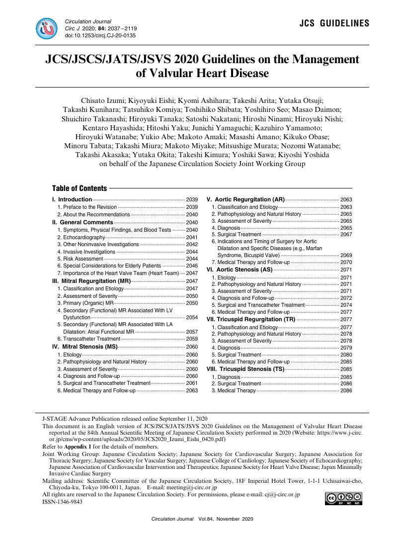
2020 Volume 84 Issue 11 Pages 2037-2119
- Published: October 23, 2020 Received: - Released on J-STAGE: October 23, 2020 Accepted: - Advance online publication: September 11, 2020 Revised: -
(compatible with EndNote, Reference Manager, ProCite, RefWorks)
(compatible with BibDesk, LaTeX)


